i


The circulatory system


    Blood vessels – arteries, veins and capillaries


    Blood vessels are tubes that carry blood in your body. There are three kinds: arteries, veins, and capillaries.


    Arteries


    Arteries take blood away from the heart after it is pumped. The blood is under high pressure. The arteries have thick and elastic muscle walls to handle this pressure. The elastic walls also let the blood pulse when your heart beats.


    Arteries:
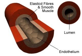
     

    • Carry blood away from the heart.
    • Have thick walls composed largely of muscle and elastic tissue.
    • Have small passageways for blood (internal lumen).
    • Contain blood under high pressure.


    This image is a diagram illustrating the cross-section of an artery, highlighting its various layers and components.


    Veins


    Veins bring blood back to the heart. The blood is under low pressure because some of it is lost as it goes around your body. The veins have thin and less elastic muscle walls than arteries. The veins have one-way valves to stop the blood from going backwards.
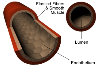

    This image is a diagram illustrating the cross-section of a vein, highlighting its various layers and components.

    Veins:


    • Carries blood back to the heart.
    • Have thin walls.
    • Have larger passageways for blood (internal lumen).
    • Contain blood under low pressure.
    • Contain valves which stop the blood flowing the wrong way


    Capillaries


    Capillaries are very small blood vessels that go into every tissue in your body. They carry oxygen and glucose for respiration to your cells and take away carbon dioxide and other wastes. They have very thin walls to let these substances move in and out of your cells by diffusion. Capillaries connect your arteries and veins.
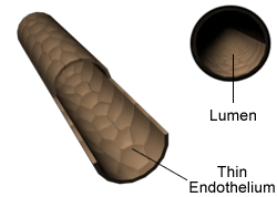
     

    This image is a diagram illustrating the cross-section of a capillary, highlighting its various layers and components.
  •  

    Capillaries:


    • Found in the muscles and lungs.
    • Walls are one cell thick to allow substances to pass over.
    • Very low blood pressure.
    • Where gas exchange takes place - oxygen passes through the capillary wall and into the tissues, while carbon dioxide passes from the tissues into the blood.


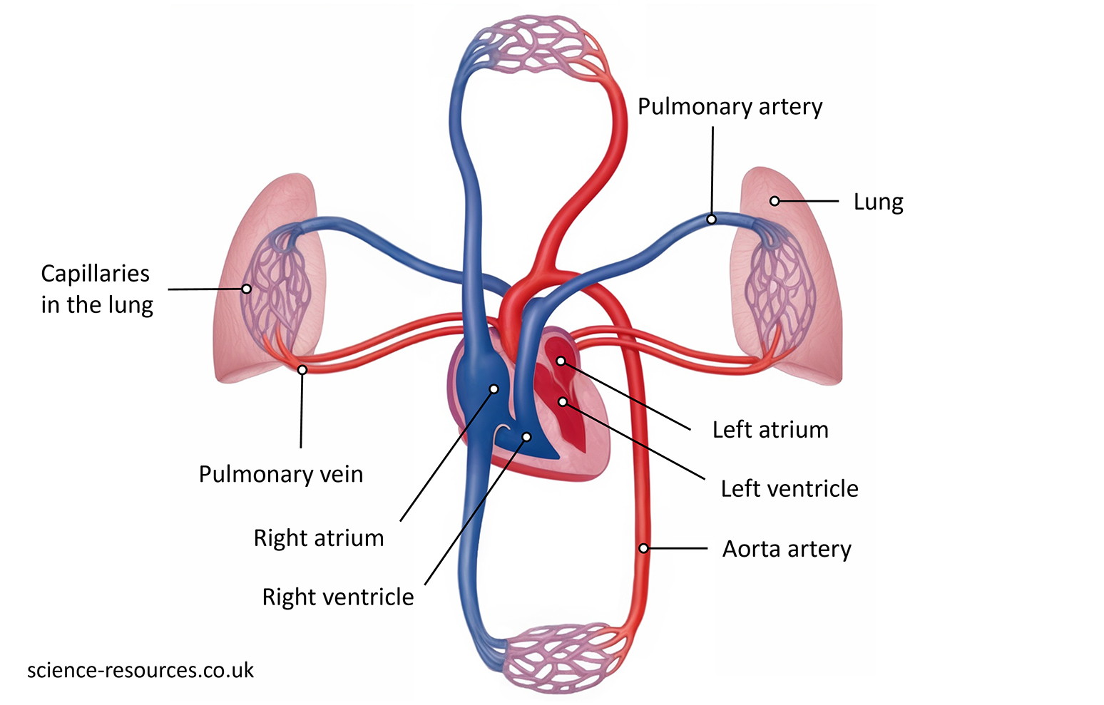
    This image is a detailed diagram of the human heart and its connection to the lungs, showcasing various parts and their names.


    The heart with the arteries, veins and capillaries


    Heart


    The heart is a powerful muscle that pumps blood to your lungs and the rest of your body. It has four sections called chambers.


    This image is a detailed diagram of the human heart, showcasing its various parts and the flow of oxygenated and deoxygenated blood.


    The image (above) shows how blood moves in the four chambers of the heart. The heart’s wall is thick and strong. The upper chambers are the atria (right and left) and the lower chambers are the ventricles (right and left). The atria receive blood and send it to the ventricles. The ventricles then push the blood to the body. The ventricles look bigger, but the chambers are all the same size. The ventricles have more muscle because they need to pump blood farther than the atria.


    Summary:


    • Blood vessels are tubes that carry blood in your body. There are three kinds: arteries, veins, and capillaries.
    • The heart is a muscle that pushes blood through your blood vessels. It makes your circulatory system work.
    • The circulatory system is the heart and all the blood vessels. It delivers cells and substances to every part of your body.


    © 2012 science-resources.co.uk. All rights reserved | Design by W3layouts